


- Description
| Application | Conditions | Recommended Concentration |
| Immunofluorescence, Extracellular | Fixed 4% paraformaldehyde | 1 µg/ml |
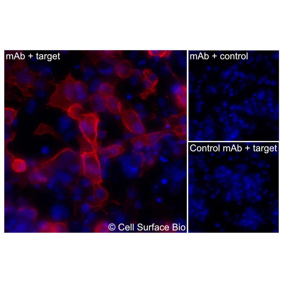 |
(Left) COS-7 cells transiently transfected human CD62L were stained with CD62L Monoclonal Antibody (CSB0223) followed by AlexaFluor 647 anti- IgG secondary antibody (red) and DAPI (blue). (Top right) COS-7 cells transiently transfected with an empty control vector stained with CD62L Monoclonal Antibody. (Bottom right) Isotype control: COS-7 cells transfected with human CD62L and stained with control MAb. | |
| Application | Conditions | Recommended Concentration |
| Immunofluorescence, Intracellular | Fixed 4% paraformaldehyde, Permeabilized 0.1% Triton X-100 | 1 µg/ml |
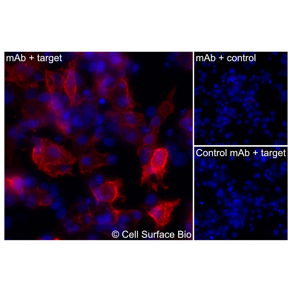 |
(Left) COS-7 cells transiently transfected human CD62L were permeabilized and stained with CD62L Monoclonal Antibody (CSB0223) followed by AlexaFluor 647 anti- IgG secondary antibody (red) and DAPI (blue). (Top right) COS-7 cells transiently transfected with an empty control vector stained with CD62L Monoclonal Antibody. (Bottom right) Isotype control: COS-7 cells transfected with human {{target} and stained with control MAb. | |
| Application | Conditions | Recommended Concentration |
| Flow Cytometry, Extracellular | Live, Unpermeabilized | 1 µg/ml |
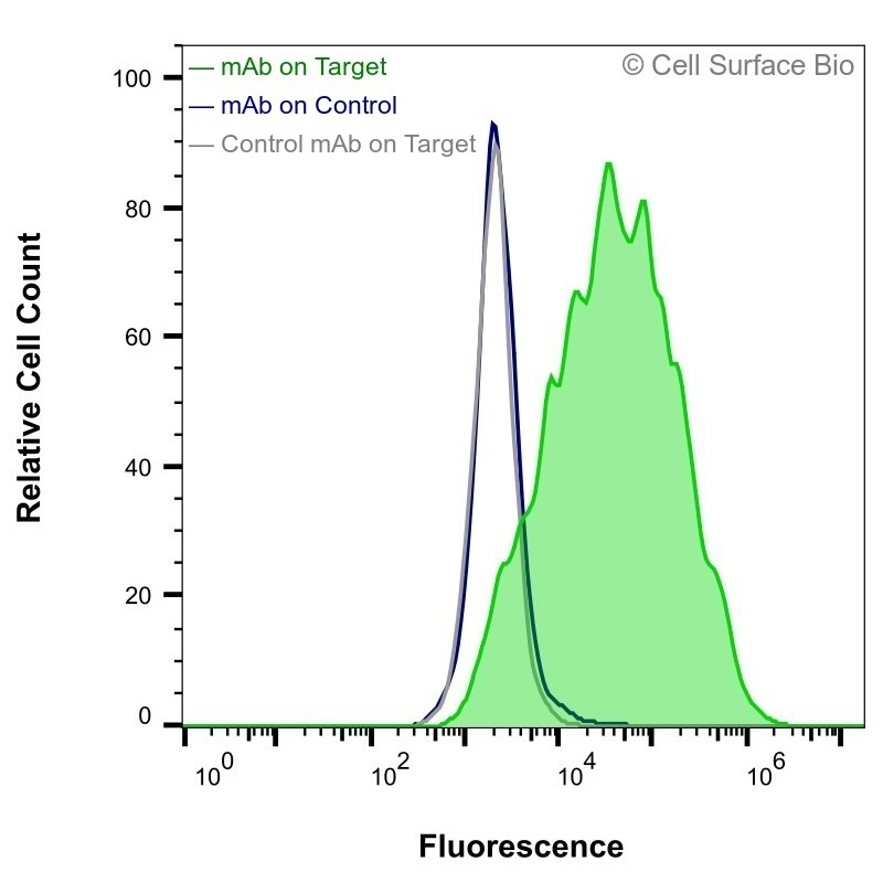 |
HEK-F cells transiently transfected with human CD62L were stained with CD62L Monoclonal Antibody (CSB0223) (green) or isotype control antibody (gray), followed by AlexaFluor 647- conjugated anti- IgG secondary antibody. HEK-F cells transiently transfected with an empty control vector were also stained with CD62L Monoclonal Antibody (CSB0223) (blue). | |
| Application | Conditions | Recommended Concentration |
| Membrane Proteome Specificity | ||
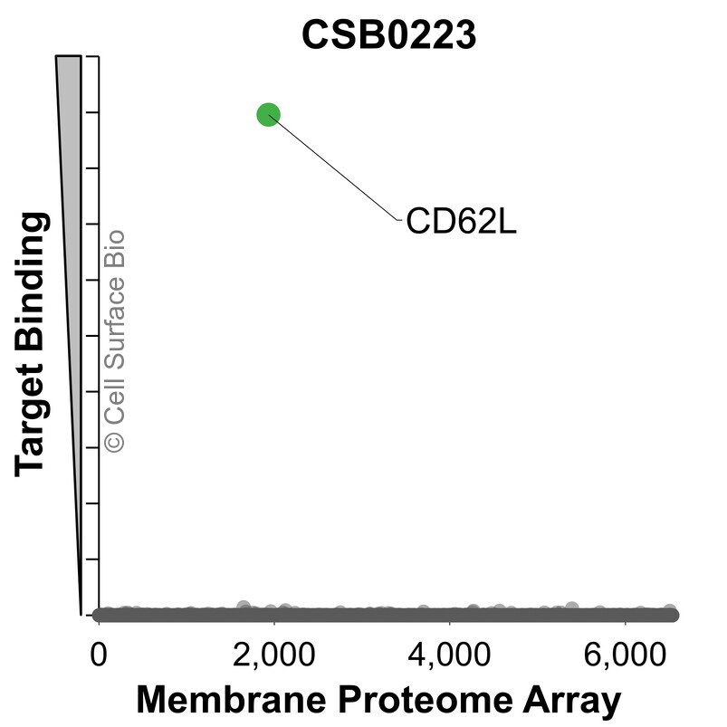 |
The specificity of CD62L Monoclonal Antibody (CSB0223) was tested on the Membrane Proteome Array™ and shown to be specific for human CD62L. The Membrane Proteome Array™ contains 6,000 different human membrane proteins, each expressed in unfixed Human cells to ensure native conformation and post-translational modifications. The Membrane Proteome Array™ represents the industry standard for determining the binding specificity of antibodies and other protein ligands. | |
CSB VeRSaMAbs:
We produce and validate all our monoclonal antibodies in-house so that you can confidently choose the right antibody for your assay. All antibodies are VALIDATED for common applications, RECOMBINANT for batch-to-batch consistency, and SPECIFIC in binding to only your target.
| Target | CD62L |
| Target Aliases | CD62L, LAM-1, LECAM1, LNHR, selectin L |
| Isotype | Mouse IgG2a |
| Species Reactivity | Human, Greater Horseshoe Bat, Egyptian Fruit Bat , Pig (+/-) |
| Concentration | 1 mg/ml |
Stable for 12 months from date of receipt when stored at 4°C. Avoid repeated freeze-thaw cycles.
L-selectin (SELL) is a type I transmembrane glycoprotein and a member of the selectin family of cell adhesion molecules. It is expressed on most leukocytes, including naïve T cells, B cells, neutrophils, and monocytes, where it mediates leukocyte rolling and homing to secondary lymphoid tissues through binding to carbohydrate ligands on endothelial cells. CD62L is a marker for T cell activation/differentiation state. The extracellular region contains a C-type lectin domain, an epidermal growth factor-like domain, and a series of short consensus repeat units, while the cytoplasmic tail contributes to intracellular signaling and shedding regulation. Proteolytic cleavage of the extracellular domain regulates its surface expression and leukocyte trafficking.