


- Description
| Application | Conditions | Recommended Concentration |
| Flow Cytometry, Extracellular | Live, Unpermeabilized | 1 µg/ml |
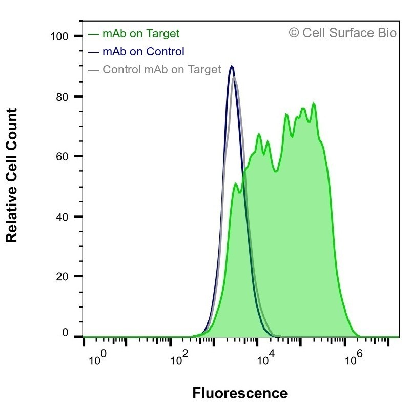 |
HEK-293F cells transiently transfected with CD16 were stained with CD16 Monoclonal Antibody (CSB0156) (green) or isotype control antibody (gray), followed by AlexaFluor 647- conjugated anti- IgG secondary antibody. HEK-293F cells transiently transfected with an empty control vector were also stained with CD16 Monoclonal Antibody (CSB0156) (blue). | |
| Application | Conditions | Recommended Concentration |
| Immunofluorescence, Extracellular | Fixed 4% paraformaldehyde | 1 µg/ml |
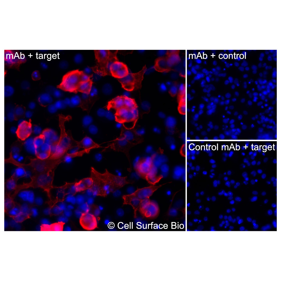 |
(Left) COS-7 cells transiently transfected CD16 were stained with CD16 Monoclonal Antibody (CSB0156) followed by AlexaFluor 647 anti- IgG secondary antibody (red) and DAPI (blue). (Top right) COS-7 cells transiently transfected with an empty control vector stained with CD16 Monoclonal Antibody. (Bottom right) Isotype control: COS-7 cells transfected with CD16 and stained with control MAb. | |
| Application | Conditions | Recommended Concentration |
| Immunofluorescence, Intracellular | Fixed 4% paraformaldehyde, Permeabilized 0.1% Triton X-100 | 1 µg/ml |
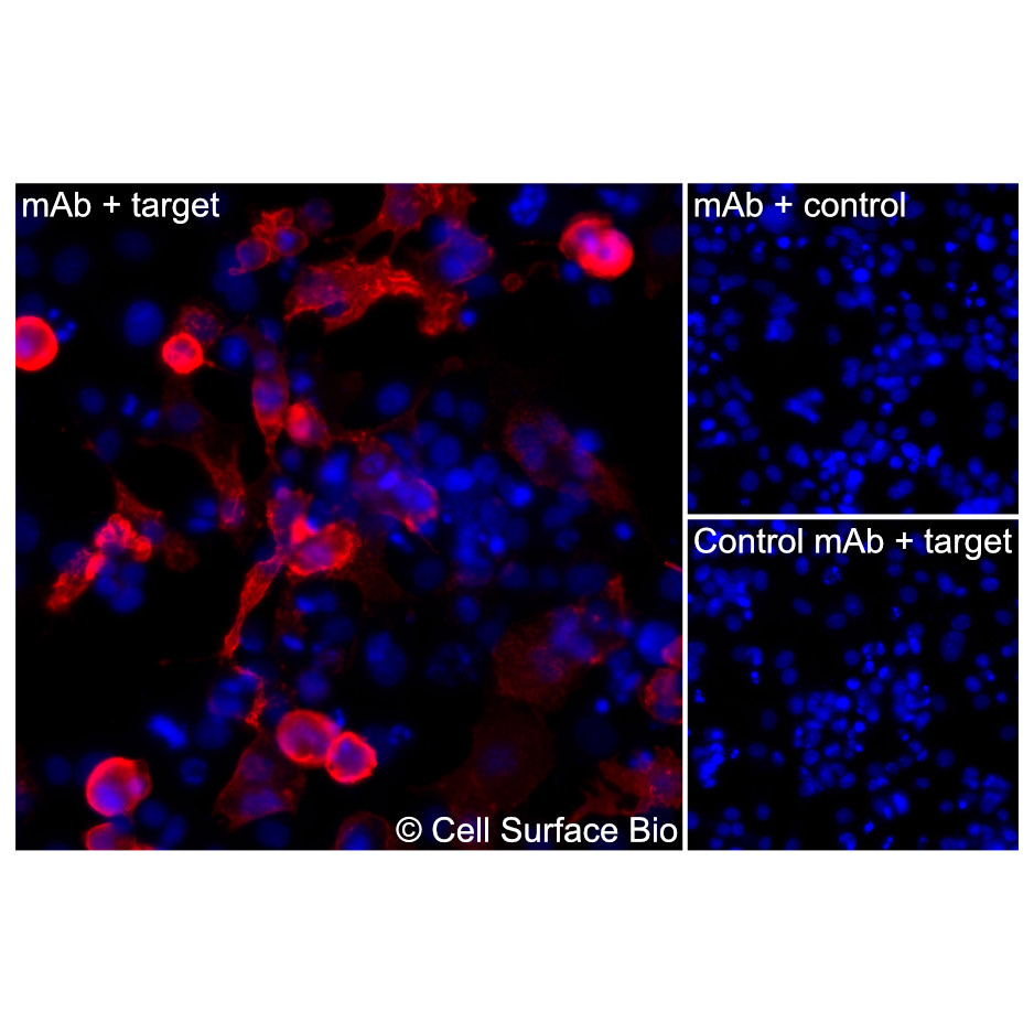 |
(Left) COS-7 cells transiently transfected CD16 were permeabilized and stained with CD16 Monoclonal Antibody (CSB0156) followed by AlexaFluor 647 anti- IgG secondary antibody (red) and DAPI (blue). (Top right) COS-7 cells transiently transfected with an empty control vector stained with CD16 Monoclonal Antibody. (Bottom right) Isotype control: COS-7 cells transfected with {{target} and stained with control MAb. | |
| Application | Conditions | Recommended Concentration |
| Membrane Proteome Specificity | ||
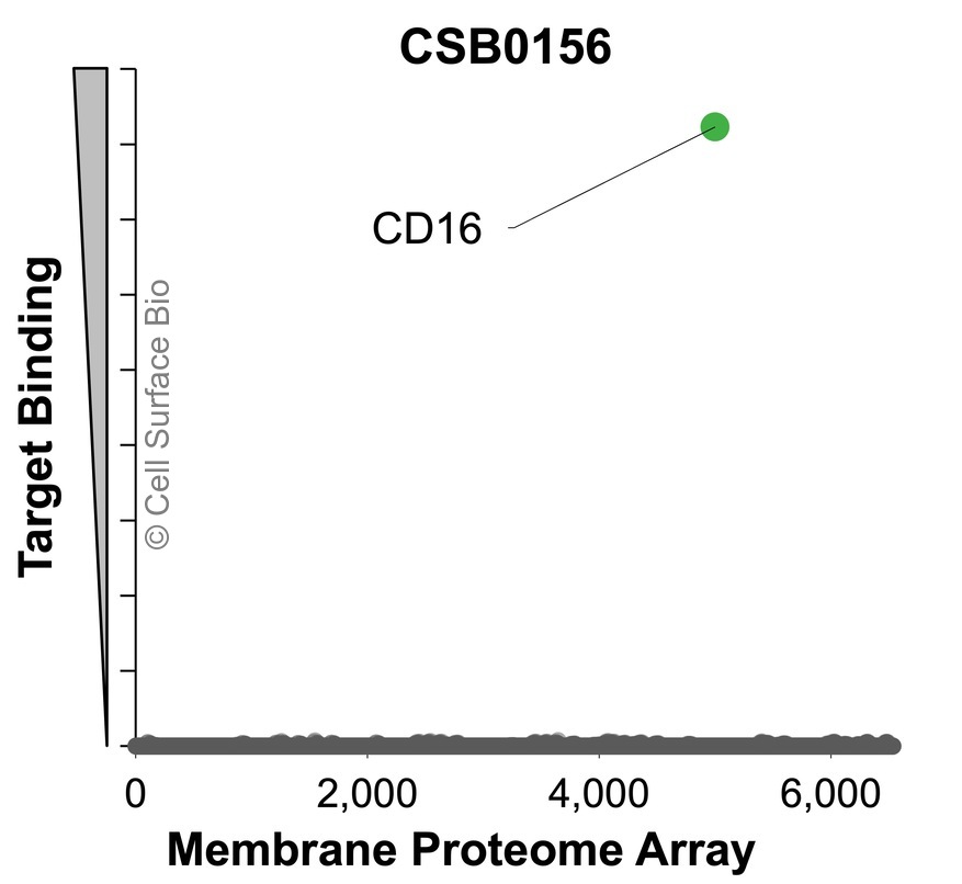 |
The specificity of CD16 Monoclonal Antibody (CSB0156) was tested on the Membrane Proteome Array™ and shown to be specific for CD16. The Membrane Proteome Array™ contains 6,000 different human membrane proteins, each expressed in unfixed avian cells to ensure native conformation and post-translational modifications. The Membrane Proteome Array™ represents the industry standard for determining the binding specificity of antibodies and other protein ligands. | |
CSB VeRSaMAbs:
We produce and validate all our monoclonal antibodies in-house so that you can confidently choose the right antibody for your assay. All antibodies are VALIDATED for common applications, RECOMBINANT for batch-to-batch consistency, and SPECIFIC in binding to only your target.
| Target | CD16 |
| Target Aliases | CD16a: FGCR3A, Fc Gamma Receptor IIIa, FcgammaRIIIa, FcGRIIIA, CD16a, FCGR3, CD16 CD16b: FCGR3B, Fc Gamma Receptor IIIb, FcRIIIb, FcgammaRIIIb, CD16b, FCGR3, CD16, FCG3 |
| Isotype | IgG2a |
| Species Reactivity | Human (Others untested) |
| Concentration | 1 mg/ml |
Stable for 12 months from date of receipt when stored at 4°C. Avoid repeated freeze-thaw cycles.
CD16 refers to two closely related receptors, FCGR3A and FCGR3B, both members of the immunoglobulin superfamily that bind the Fc portion of IgG antibodies with low affinity. These proteins contain extracellular immunoglobulin-like domains and differ in their membrane anchoring: FCGR3A is a transmembrane protein expressed on natural killer (NK) cells, macrophages, and subsets of T cells, while FCGR3B is GPI-anchored and primarily found on neutrophils. CD16 mediates antibody-dependent cellular cytotoxicity (ADCC) and other immune effector functions through IgG binding. These receptors play a central role in linking the adaptive humoral response to innate immune mechanisms.