| Application |
Conditions |
Recommended Concentration |
| Membrane Proteome Specificity |
|
|
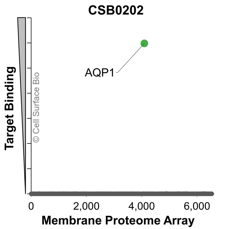 |
The specificity of AQP1 Monoclonal Antibody (CSB0202) was tested on the Membrane Proteome Array™ and shown to be specific for AQP1. The Membrane Proteome Array™ contains 6,000 different human membrane proteins, each expressed in unfixed avian cells to ensure native conformation and post-translational modifications. The Membrane Proteome Array™ represents the industry standard for determining the binding specificity of antibodies and other protein ligands. |
| Application |
Conditions |
Recommended Concentration |
| Flow Cytometry, Extracellular |
Live, Unpermeabilized |
1 µg/ml |
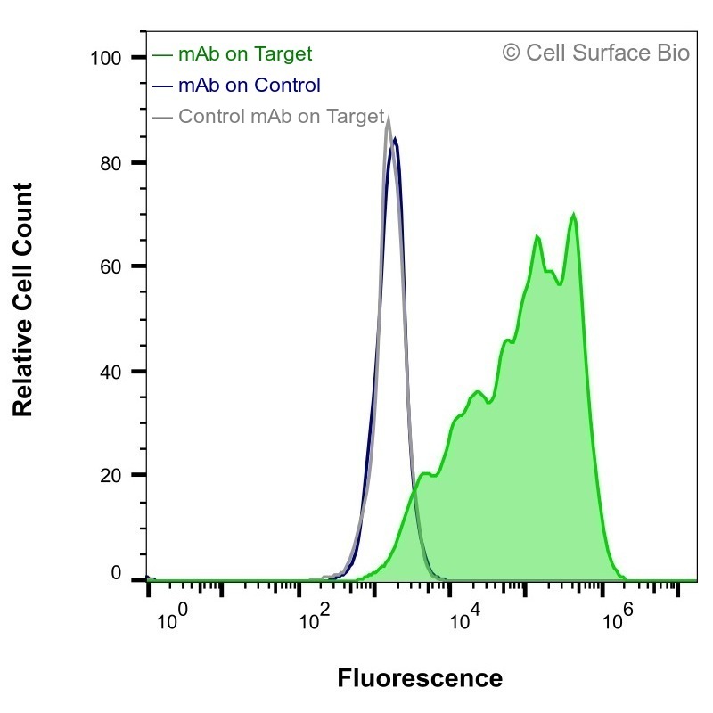 |
JS-1 cells transiently transfected with AQP1 were stained with AQP1 Monoclonal Antibody (CSB0202) (green) or isotype control antibody (gray), followed by AlexaFluor 647- conjugated anti- IgG secondary antibody. JS-1 cells transiently transfected with an empty control vector were also stained with AQP1 Monoclonal Antibody (CSB0202) (blue). |
| Application |
Conditions |
Recommended Concentration |
| Immunofluorescence, Intracellular |
Fixed 4% paraformaldehyde, Permeabilized 0.1% Triton X-100 |
1 µg/ml |
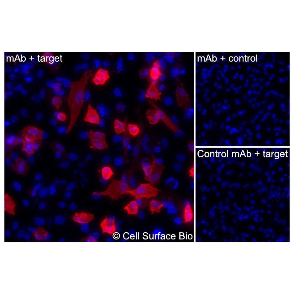 |
(Left) JS-1 cells transiently transfected AQP1 were permeabilized and stained with AQP1 Monoclonal Antibody (CSB0202) followed by AlexaFluor 647 anti- IgG secondary antibody (red) and DAPI (blue). (Top right) JS-1 cells transiently transfected with an empty control vector stained with AQP1 Monoclonal Antibody. (Bottom right) Isotype control: JS-1 cells transfected with {{target} and stained with control MAb. |
| Application |
Conditions |
Recommended Concentration |
| Immunofluorescence, Extracellular |
Fixed 4% paraformaldehyde |
1 µg/ml |
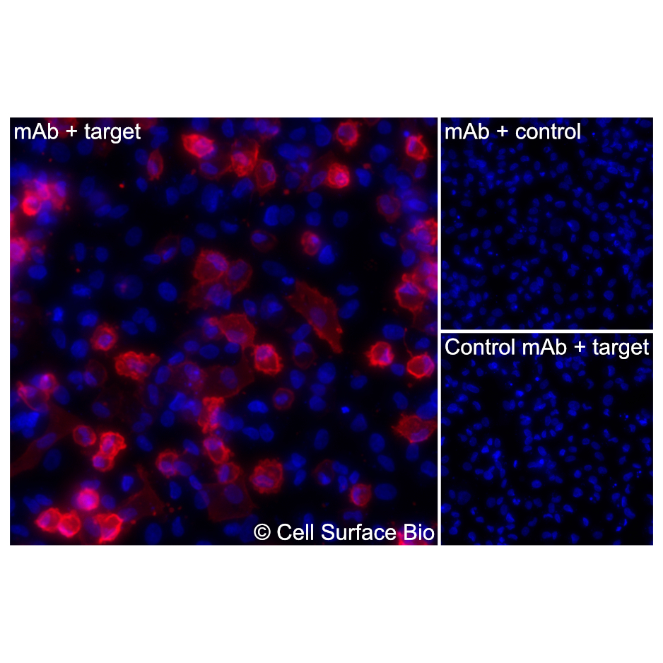 |
(Left) JS-1 cells transiently transfected AQP1 were stained with AQP1 Monoclonal Antibody (CSB0202) followed by AlexaFluor 647 anti- IgG secondary antibody (red) and DAPI (blue). (Top right) JS-1 cells transiently transfected with an empty control vector stained with AQP1 Monoclonal Antibody. (Bottom right) Isotype control: JS-1 cells transfected with AQP1 and stained with control MAb. |
CSB VeRSaMAbs:
We produce and validate all our monoclonal antibodies in-house so that you can confidently choose the right antibody for your assay. All antibodies are VALIDATED for common applications, RECOMBINANT for batch-to-batch consistency, and SPECIFIC in binding to only your target.
| Target |
AQP1 |
| Target Aliases |
Aquaporin 1 (Colton Blood Group), CHIP28,
Aquaporin 1 (Channel-Forming Integral Protein, 28kDa, CO Blood Group), Urine Water Channel,
Aquaporin-CHIP |
| Isotype |
IgG2a |
| Species Reactivity |
Human (Others untested) |
| Concentration |
1 mg/ml |
Stable for 12 months from date of receipt when stored at 4°C. Avoid repeated freeze-thaw cycles.
Aquaporin 1 is a member of the aquaporin family of integral membrane channel proteins that facilitate selective water transport across cell membranes. The protein forms homotetramers in the membrane, with each monomer containing six transmembrane helices and two conserved NPA (Asn-Pro-Ala) motifs that contribute to the water-selective pore. AQP1 enables rapid osmotic water movement and is expressed in tissues including kidney, lung, eye, and vascular endothelium. Subcellular localization studies identify AQP1 at the plasma membrane.